THE ACCUBLATE LINE OF PRODUCTS
Accublate™ is our core ablation simulation technology. We provide this technology to physicians in part by making partnership agreements with ablation system manufacturers, and by developing and offering ablation guidance software directly.
These three products are in development:
Accublate Charts is the simplest of our products, it does not incorporate a simulation technology, but it supports physicians in percutaneous RF and Microwave ablations by displaying the manufacturer’s ablation footprint overlaid to intra-procedural CT images for several supported ablation systems.
Accublate RF Sim incorporates the simulation technology which predicts the ablated volume based on a number of factors, including the true applied power, the actual shape of the deployed RF tines as recognized from images, and patient specific aspects which include the heat-sink effect of vasculature.
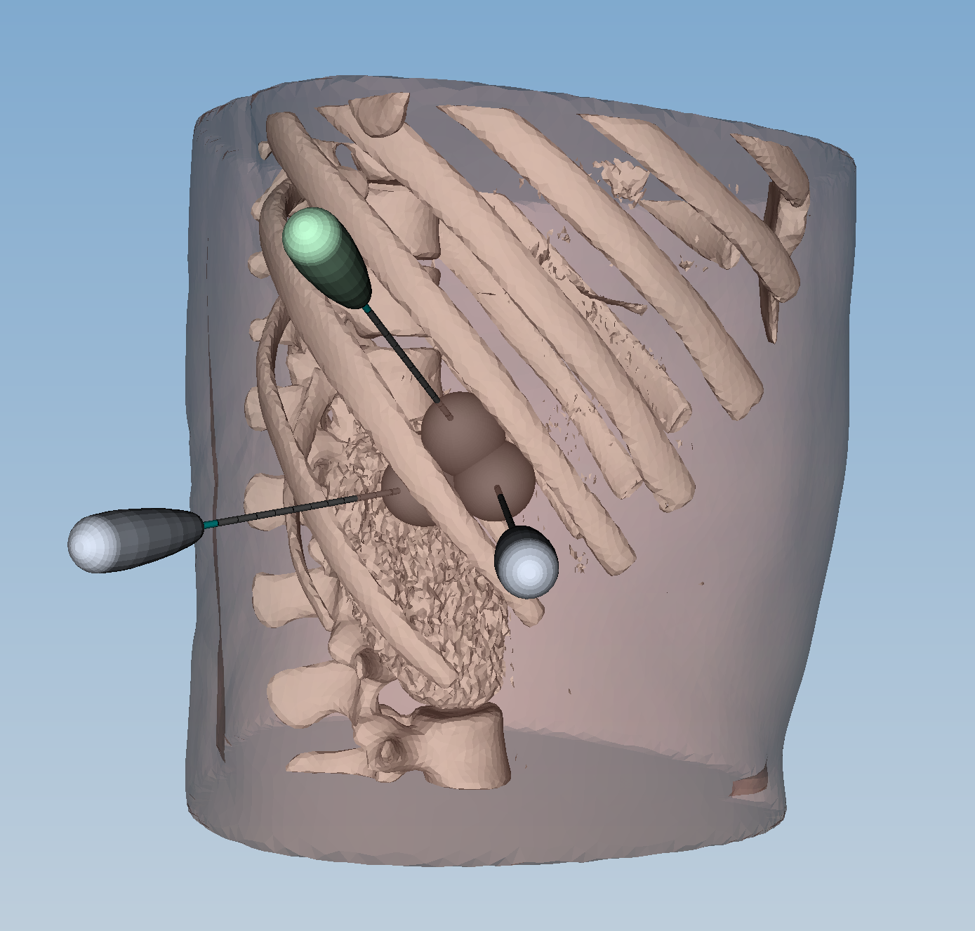
Accublate MW Sim similarly to Accublate RF Sim, incorporates our simulation technology for predicting the ablation volume based on procedure and patient specific factors. Accublate MW Sim incorporates support for multiple ablation probes activated concurrently.
NE Scientific is seeking regulatory approvals for the Accublate line, the products are not currently commercially available. Accublate RF Sim has been employed in a clinical prospective clinical trial for guidance of RF liver cancer ablation. Recruitment for the trial has terminated and interim results indicate an excellent efficacy in reducing local recurrence. Please visit our Clinical Trials page for more information.

ACCUBLATE IN USE
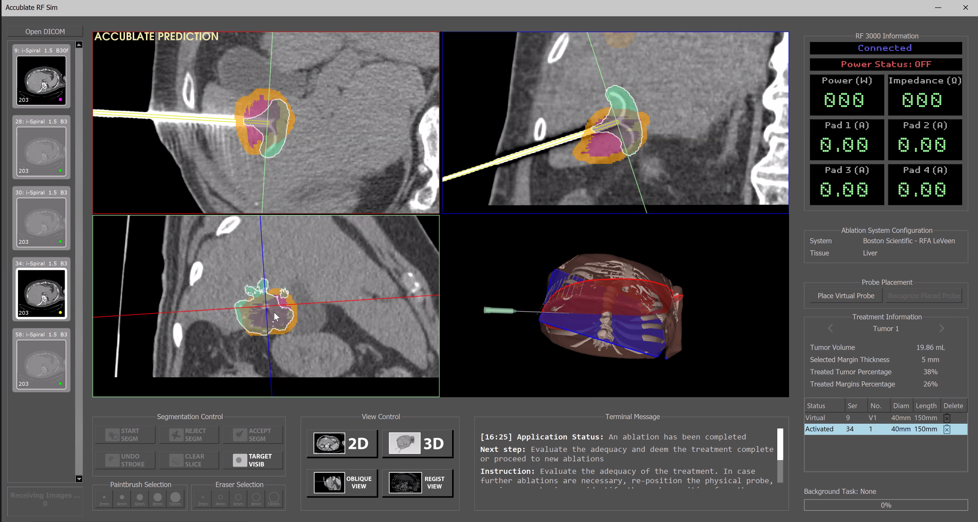
GRAPHICAL USER INTERFACE: Accubalte has a easy to use graphical user interface and a workflow that integrates with the current standard clinical practice without requiring changes to setup or images that acquired. The learnign curve is modest ,the sopport of the software simplifies the evaluation of adequacy, and the mental burden on the physician is reduced.
Malignant tissues are segmented manually at the start of the procedure, and indicated in red. Margins are built automatically by the application and indicated in yellow. Abalted tissues are indicated in green.
Evaluation of the status of tissues can occur in the traditional axial, coronal, or saggital views, or in multplanar views automatically setup by Accublate, with slicing palnes passing by the shaft of the probe.

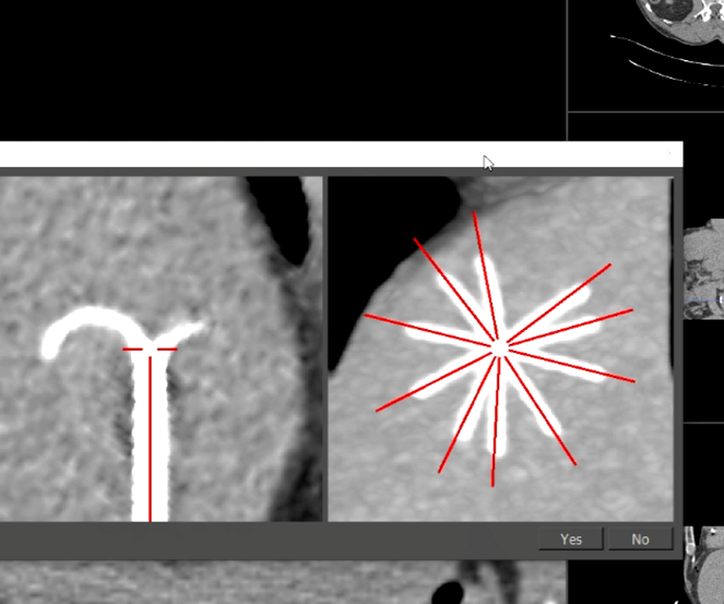
PROBE GEOMETRY RECOGNITION: in RF applications often abaltion probes deploy tinaes in the tissues in order to attain a larger abaltion volume. Tines are thin shape-meomory filaments that when subject to the stiffness of tissues might deploy in irregular shapes compared to what expected. Accubalte analyzes the image to recognize the position and shape of each single tine, building on the flight a model of the deployed probe and tines, and providing a prediction of the abalted volume that reflects the actual geoemtry of the probe as deployed in the tissues.


PROBE GEOMETRY RECOGNITION: in RF applications often abaltion probes deploy tinaes in the tissues in order to attain a larger abaltion volume. Tines are thin shape-meomory filaments that when subject to the stiffness of tissues might deploy in irregular shapes compared to what expected. Accubalte analyzes the image to recognize the position and shape of each single tine, building on the flight a model of the deployed probe and tines, and providing a prediction of the abalted volume that reflects the actual geoemtry of the probe as deployed in the tissues.
MULTIPLE OVERLAPPING ABLATIONS: Accublate supports the abaltion of larger tumors by overlapping abaltions by accumulating the effect of each single abaltion in a tissue damage map which is updated at each new abaltion. The physician does not need to relay on mental maps of which part of the tumor has been ablated and which not to any point in the procedure: the application shows the current status of tissues, as resulting from all the abaltions performed.

